Loculated Pleural Effusion - Loculated pleural effusion | Radiology Case | Radiopaedia.org / We studied the value of transca …
bymanamcolasante-
0
Loculated Pleural Effusion - Loculated pleural effusion | Radiology Case | Radiopaedia.org / We studied the value of transca …. Pleural effusion occurs when too much fluid collects in the pleural space (the space between the two layers of the pleura). Treatment may fail if the catheter is not placed optimally within the loculation or if the fluid is hemorrhagic or fibrinous. The latter are more likely to change with patient positioning 12. Strange or atypical configurations of pleural fluid can be due to either adhesions (i.e. It is commonly known as water on the lungs.
We studied the value of transca … Pleural effusion that is confined to one or more fixed pockets in the pleural space. Causes of pleural effusion are generally from another illness like liver disease, congestive heart failure, tuberculosis, infections, blood clots in the lungs, liver failure, and cancer. Strange or atypical configurations of pleural fluid can be due to either adhesions (i.e. Pleural effusion occurs when too much fluid collects in the pleural space (the space between the two layers of the pleura).
Loculated pleural effusion | Image | Radiopaedia.org from images.radiopaedia.org Surgical thoracostomy tube placement and radiologically guided catheter drainage are standard therapy for loculated pleural fluid collections. Pleural effusion (basic) large unilateral pleural effusion; May 25, 2021 · the aetiology of the pleural effusion determines other signs and symptoms. Pleural effusion in other conditions classified elsewhere secondary pleural effusion ; The latter are more likely to change with patient positioning 12. Blunting of the lateral costophrenic angle usually requires about 175 ml but may take as much as 500 ml. Strange or atypical configurations of pleural fluid can be due to either adhesions (i.e. If your doctor suspects a malignant pleural effusion, the next step is usually a thoracentesis, a procedure in which a needle is inserted through the chest wall into the pleural space to get a sample of the fluid.
Strange or atypical configurations of pleural fluid can be due to either adhesions (i.e.
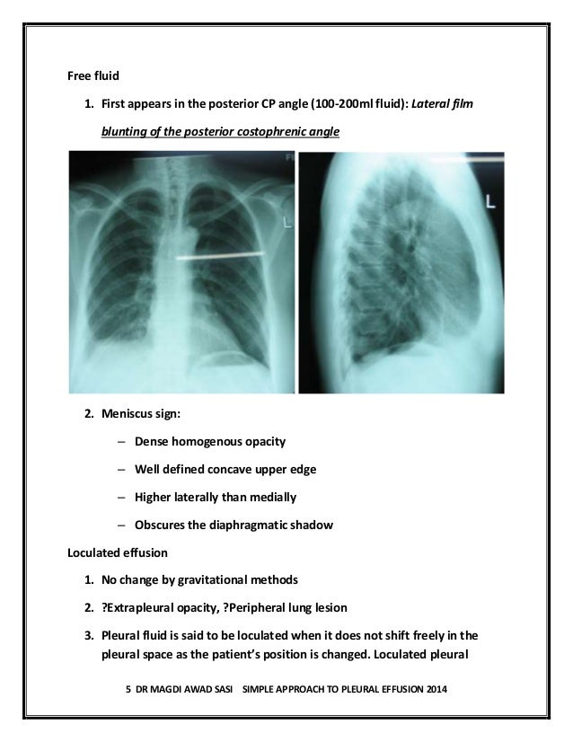
Blunting of the lateral costophrenic angle usually requires about 175 ml but may take as much as 500 ml. Loculated effusion) or underlying atelectasis. Obliteration of left costophrenic angle with a wide pleural based dome shaped opacity projecting into the lung noted tracking along the cp angle and lateral chest wall suggestive of loculated pleural effusion, however the possibility of empyema can not be ruled out completely. If your doctor suspects a malignant pleural effusion, the next step is usually a thoracentesis, a procedure in which a needle is inserted through the chest wall into the pleural space to get a sample of the fluid. Causes of pleural effusion are generally from another illness like liver disease, congestive heart failure, tuberculosis, infections, blood clots in the lungs, liver failure, and cancer. Surgical thoracostomy tube placement and radiologically guided catheter drainage are standard therapy for loculated pleural fluid collections. Pleural effusion in other conditions classified elsewhere secondary pleural effusion ; The latter are more likely to change with patient positioning 12. Pleural effusion (basic) large unilateral pleural effusion; It is commonly known as water on the lungs. Pleural effusion occurs when too much fluid collects in the pleural space (the space between the two layers of the pleura). Pleural effusion that is confined to one or more fixed pockets in the pleural space. Feb 07, 2020 · learn about pleural effusion (fluid in the lung) symptoms like shortness of breath and chest pain.
May 25, 2021 · the aetiology of the pleural effusion determines other signs and symptoms. Blunting of the lateral costophrenic angle usually requires about 175 ml but may take as much as 500 ml. Pleural effusion in other conditions classified elsewhere secondary pleural effusion ; Surgical thoracostomy tube placement and radiologically guided catheter drainage are standard therapy for loculated pleural fluid collections. Obliteration of left costophrenic angle with a wide pleural based dome shaped opacity projecting into the lung noted tracking along the cp angle and lateral chest wall suggestive of loculated pleural effusion, however the possibility of empyema can not be ruled out completely.
Pleural effusion dr magdi sasi from image.slidesharecdn.com Blunting of the lateral costophrenic angle usually requires about 175 ml but may take as much as 500 ml. We studied the value of transca … Feb 07, 2020 · learn about pleural effusion (fluid in the lung) symptoms like shortness of breath and chest pain. It is commonly known as water on the lungs. Surgical thoracostomy tube placement and radiologically guided catheter drainage are standard therapy for loculated pleural fluid collections. Pleural effusion occurs when too much fluid collects in the pleural space (the space between the two layers of the pleura). Loculated effusion) or underlying atelectasis. May 25, 2021 · the aetiology of the pleural effusion determines other signs and symptoms.
Surgical thoracostomy tube placement and radiologically guided catheter drainage are standard therapy for loculated pleural fluid collections.
It is commonly known as water on the lungs. We studied the value of transca … Loculated effusion) or underlying atelectasis. Treatment may fail if the catheter is not placed optimally within the loculation or if the fluid is hemorrhagic or fibrinous. Pleural effusion occurs when too much fluid collects in the pleural space (the space between the two layers of the pleura). Pleural effusion that is confined to one or more fixed pockets in the pleural space. Obliteration of left costophrenic angle with a wide pleural based dome shaped opacity projecting into the lung noted tracking along the cp angle and lateral chest wall suggestive of loculated pleural effusion, however the possibility of empyema can not be ruled out completely. Pleural effusion in other conditions classified elsewhere secondary pleural effusion ; The latter are more likely to change with patient positioning 12. Surgical thoracostomy tube placement and radiologically guided catheter drainage are standard therapy for loculated pleural fluid collections. Pleural effusion (basic) large unilateral pleural effusion; Feb 07, 2020 · learn about pleural effusion (fluid in the lung) symptoms like shortness of breath and chest pain. If your doctor suspects a malignant pleural effusion, the next step is usually a thoracentesis, a procedure in which a needle is inserted through the chest wall into the pleural space to get a sample of the fluid.
Treatment may fail if the catheter is not placed optimally within the loculation or if the fluid is hemorrhagic or fibrinous. Blunting of the lateral costophrenic angle usually requires about 175 ml but may take as much as 500 ml. Feb 07, 2020 · learn about pleural effusion (fluid in the lung) symptoms like shortness of breath and chest pain. Pleural effusion occurs when too much fluid collects in the pleural space (the space between the two layers of the pleura). We studied the value of transca …
Calcinosis in CREST syndrome | Image | Radiopaedia.org from images.radiopaedia.org It is commonly known as water on the lungs. We studied the value of transca … Pleural effusion that is confined to one or more fixed pockets in the pleural space. Loculated effusion) or underlying atelectasis. May 25, 2021 · the aetiology of the pleural effusion determines other signs and symptoms. The latter are more likely to change with patient positioning 12. Treatment may fail if the catheter is not placed optimally within the loculation or if the fluid is hemorrhagic or fibrinous. If your doctor suspects a malignant pleural effusion, the next step is usually a thoracentesis, a procedure in which a needle is inserted through the chest wall into the pleural space to get a sample of the fluid.
May 25, 2021 · the aetiology of the pleural effusion determines other signs and symptoms.
Pleural effusion occurs when too much fluid collects in the pleural space (the space between the two layers of the pleura). Loculated effusion) or underlying atelectasis. May 25, 2021 · the aetiology of the pleural effusion determines other signs and symptoms. Pleural effusion that is confined to one or more fixed pockets in the pleural space. It is commonly known as water on the lungs. Obliteration of left costophrenic angle with a wide pleural based dome shaped opacity projecting into the lung noted tracking along the cp angle and lateral chest wall suggestive of loculated pleural effusion, however the possibility of empyema can not be ruled out completely. Treatment may fail if the catheter is not placed optimally within the loculation or if the fluid is hemorrhagic or fibrinous. We studied the value of transca … Pleural effusion in other conditions classified elsewhere secondary pleural effusion ; Surgical thoracostomy tube placement and radiologically guided catheter drainage are standard therapy for loculated pleural fluid collections. Blunting of the lateral costophrenic angle usually requires about 175 ml but may take as much as 500 ml. Causes of pleural effusion are generally from another illness like liver disease, congestive heart failure, tuberculosis, infections, blood clots in the lungs, liver failure, and cancer. Feb 07, 2020 · learn about pleural effusion (fluid in the lung) symptoms like shortness of breath and chest pain.